Magnetic Resonance Imaging (MRI)
Syllabus: Science and Technology [GS Paper-3]

Context
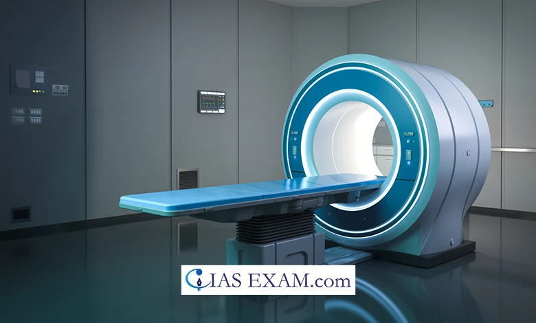
Magnetic Resonance Imaging (MRI) is a sophisticated medical imaging technique that has revolutionised the field of diagnostic medicine. It provides a non-invasive way to visualise the internal structures of the body in high detail, particularly soft tissues, which are often not as clearly seen with other imaging methods like X-rays or CT scans.
How MRI Works
- MRI is built on the concept of nuclear magnetic resonance, where nuclei in a magnetic field absorb and emit electromagnetic radiation.
- In an MRI scan, the body is placed in a strong magnetic field, aligning hydrogen protons in water and fat molecules.
- Radiofrequency pulses are used to create signals from these aligned protons, which are then used to generate detailed images of the body’s internal structures.
Components of an MRI System
An MRI system consists of several key components:
- Magnet: The most important part of the MRI machinery is the magnet; instead of using any other magnets in their machines, superconducting magnets are used, known for their huge and stable magnetic field.
- Shim Coils: These serve in order to restore the magnetic field during the occurrence of its inhomogeneities to provide uniformity. I can always use an extra hand with my homework assignments, and the efficient online writing community helps me bridge the gap.
- Gradient Coils: These coils produce a variable magnetic field over the region of the area under the region of interest, thereby, permitting spatial modulation of the MRI signals.
- RF Coils: The radiofrequency coil would be used to send radio waves into the body and afterward receive the signal.
MRI Sequences and Imaging
MRI sequences are the specific settings and protocols used during the scan to create different types of images. Some common sequences include:
- T1-Weighted Imaging: Convenient for both anatomy normalisation and for tissue localization and delineation.
- T2-Weighted Imaging: A bright colour is used for areas that are filled with liquids, and the result is showing edema and inflammation.
- Diffusion-Weighted Imaging (DWI): It can accurately detect the movement of water molecules and it can also detect the alterations in tissue structure.
Applications of MRI
MRI has a wide range of applications in medical diagnosis and treatment planning:
- Neuroimaging: Thanks to its unique ability to produce images of the brain and spinal cord, MRI is particularly useful as a diagnostic tool for knowing a stroked condition, a brain tumour, or multiple sclerosis.
- Musculoskeletal Imaging: It is a medical technology which is indispensable in terms of assessing joints, muscles and bones against injuries and degenerative diseases.
- Cardiac MRI: Equipment helps to get the highly detailed images of the heart and the veins. It is used by physicians when they want to find out chronic cardiovascular diseases in patients.
- Oncology: MRI helps one to find out the cancer stage by revealing the degree of involvement due to the tumour infiltration.
Advantages of MRI
- No Ionizing Radiation: Unlike X-rays or CT scans, MRI does not use ionising radiation, making it a safer option, especially for repeated use.
- High Contrast Resolution: MRI offers superior contrast between different soft tissues of the body, making it ideal for detecting abnormalities.
- Non-invasive and pain-free
- No radiation exposure
- Can be used to diagnose a wide range of medical conditions
- Can be used to monitor treatment progress
Challenges and Considerations
- Time-Consuming: MRI scans can take a long time, sometimes up to an hour or more, which can be uncomfortable for the patient.
- Noise: The operation of gradient coils can produce loud knocking sounds, which may require ear protection.
- Claustrophobia: The confined space of the MRI tunnel can cause discomfort or anxiety in some patients, though open MRI machines are available to alleviate this issue.
- Metallic Implants: Patients with certain metallic implants, pacemakers, or metal fragments cannot undergo MRI due to the strong magnetic field.
Recent Developments in MRI
- Functional MRI (fMRI) to study brain activity
- Magnetic Resonance Angiography (MRA) to study blood vessels
- Magnetic Resonance Spectroscopy (MRS) to study metabolic disorders
- High-field MRI for improved image resolution and diagnostic accuracy
Conclusion
Magnetic Resonance Imaging (MRI) is a powerful diagnostic tool that has revolutionized the field of medical imaging. Its high-resolution images, non-invasive nature, and lack of radiation exposure make it an ideal choice for a wide range of medical conditions. While it has some limitations, recent developments in MRI technology have further expanded its applications and improved its diagnostic accuracy.
Source: The Hindu
UPSC Prelims Practice Question
Q. Which of the following medical imaging techniques utilises the principle of nuclear magnetic resonance to produce detailed images of the internal structures of the human body?
a) X-ray imaging
b) Ultrasound imaging
c) Magnetic Resonance Imaging (MRI)
d) Computed Tomography (CT) scanningAns – “c”


.png)


